
Patellofemoral Joint Instability Treatments
Conservative treatment
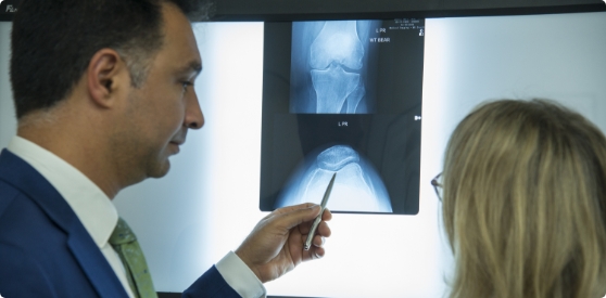
If your patella is only partially dislocated (subluxation), he may recommend non-surgical treatments, such as pain medications, rest, ice, physical therapy, knee-bracing, and orthotics. If the kneecap has been completely dislocated, the kneecap may need to be repositioned back in its proper place in the groove. This process is called closed reduction.
Surgical treatment
Surgery is sometimes needed to help return the patella to a normal tracking path when other non-surgical treatments have failed. The aim of the surgery is to realign the kneecap in the groove and to decrease the Q angle.
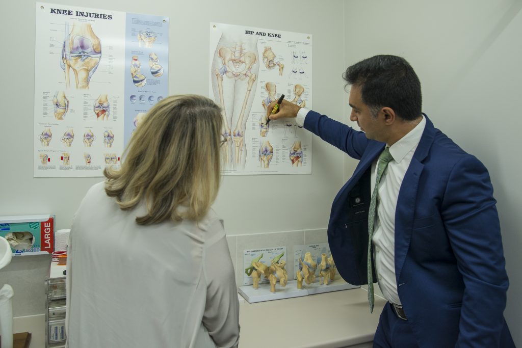
Patellar realignment surgery is broadly classified into proximal re-alignment procedures and distal re-alignment procedures.
Proximal re-alignment procedures: During this procedure, structures that limit the movements on the outside of the patella are lengthened or ligaments on the inside of the patella are shortened or reinforced with the help of ligament reconstruction.
Distal re-alignment procedures: During this procedure, the Q angle is decreased by moving the tibial tubercle towards the inner side of the knee.
The surgery is performed under sterile conditions in the operating room under spinal or general anaesthesia. Dr Khatib will make two or three small cuts around your knee. The arthroscope, a narrow tube with a tiny camera on the end is inserted through one of the incisions to view the knee joint. Specialised instruments are inserted into the joint through other small incisions. The camera attached to the arthroscope displays the image of the joint on the monitor. A sterile solution will be pumped into your knee in order to stretch the knee and provide a clear view and room for the surgeon to work. With the images from the arthroscope as a guide Dr Khatib can look for any pathology or anomaly and repair it through the other incisions with various instruments. After the evaluation is completed, a larger incision is made over the front of the knee.
Depending on your situation, a lateral retinacular release may be performed. In this procedure, the tight ligaments on the outer side of the knee are released, thus allowing the patella to sit properly in the femoral groove. A commonly performed procedure is the medial patella-femoral ligament reconstruction (MPFL), where a tendon is used to reconstruct the normal medial restrains to prevent future dislocation.
In cases where the malalignment is severe, a procedure called a tibial tubercle transfer (TTT) or Fulkerson osteotomy will be performed. In this procedure a section of bone where the patellar tendon attaches to the tibia is moved medially and sometimes downward. This bony section is then shifted and properly realigned with the patella and reattached to the tibia using screws. Once the malalignment is repaired and confirmed with arthroscopic evaluation, the incisions are closed with sutures.
Book an Appointment Today!
Postoperative care
The anterior cruciate ligament is one of the major stabilizing ligaments in the knee. It is a strong rope like structure located in the centre of the knee running from the femur to the tibia. Many activities like sport, work, mountain bike and motorcycle injuries can lead to ACL tears. Common mechanisms include knee hyperextension or pivoting on the planted knee such as when stopping and turning in netball or sideways stepping in soccer or rugby. When this ligament tears unfortunately, it does not heal and often leads to the feeling of instability in the knee. After the injury, the knee tends to swell and become painful and weight bearing may be difficult.
Risks and complications
Possible risks and complications associated with the surgery include:
- Loss of ability to extend the knee.
- Recurrent dislocations or subluxations.
- Arthrofibrosis (thick fibrous material around the joint).
Patellofemoral Joint Instability Conditions
View the conditions for Patellofemoral Joint Instability